Advanced Diagnostic Support Tool
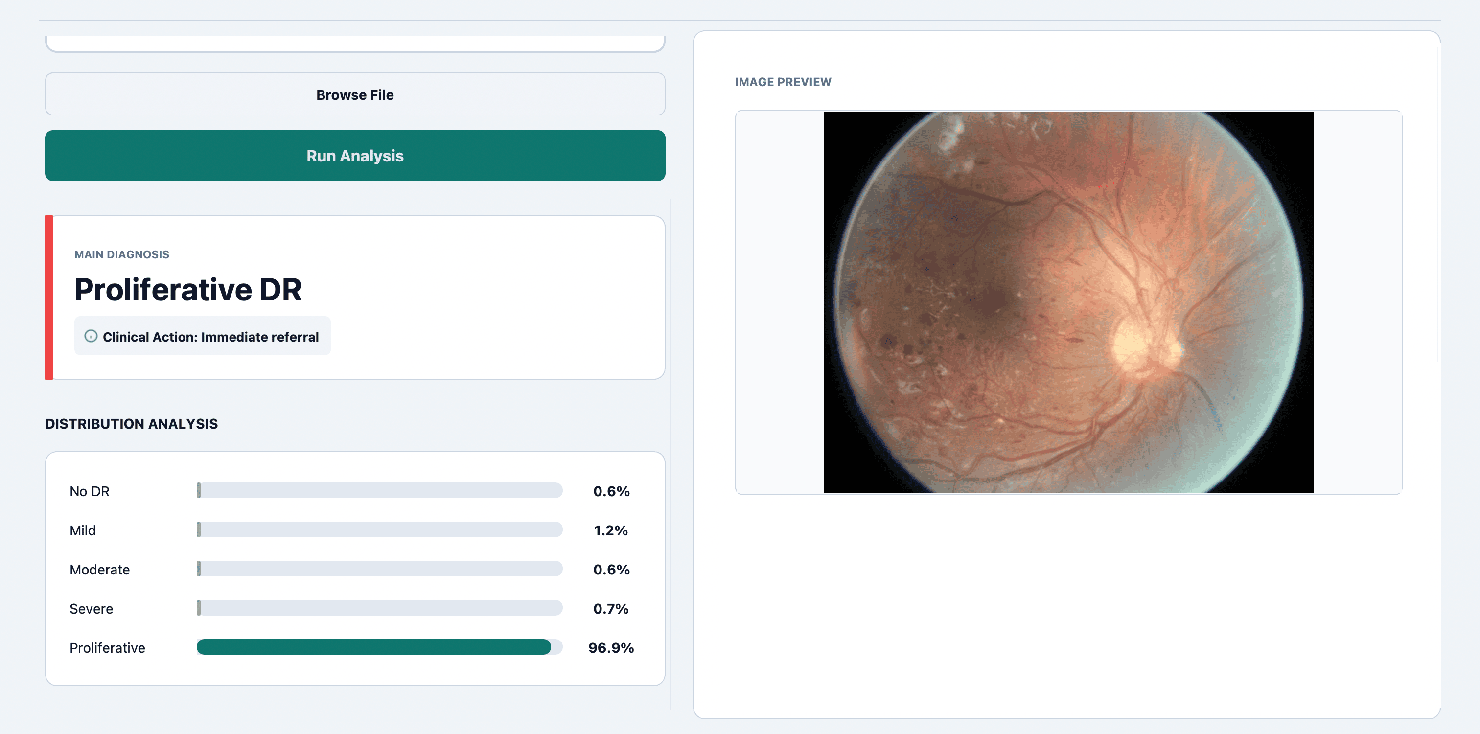
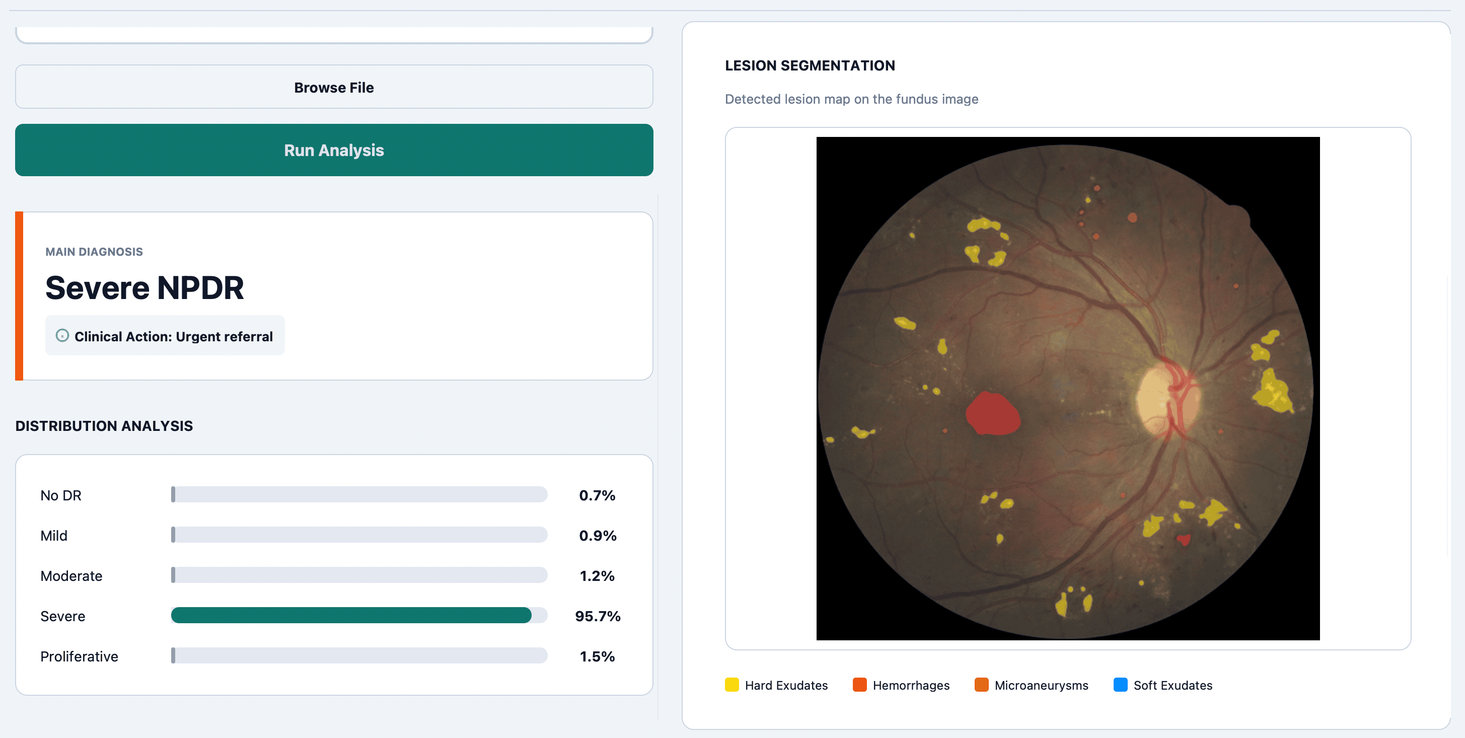
Automated Diabetic Retinopathy Detection
Empowering clinicians with a Hybrid Ensemble Model combining Swin-Base, EfficientNetV2-M, and ConvNeXt-Base. Accurately identify vascular abnormalities and grade severity in seconds.